

Case application for CT anatomy (2)
Interpret the following CT brain carefully.
Step-to-step interpretation :
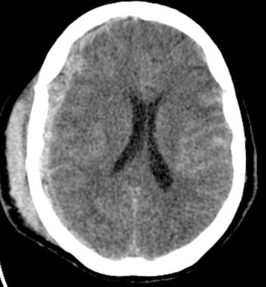
This case is a good supplementation to the previous case. This diagram is at the level of the body of lateral ventricle ( A ) also. There is a crescentic hemorrhage on the right cerebral hemisphere crossing the sutures. This is suggestive of right-sided subdural hematoma.
Apart from this, we can see a scalp hematoma which is highly suggestive of head trauma.
Owing to the space-occupying effect of the subdural hematoma, there is a mild mid-line shift. Apart from this, if we look carefully, we can see the sulcus on the left hemisphere becomes whitish, this is suggestive subarachnoid hemorrhage. In summary, there is a right-sided subdural hematoma and scalp hematoma with mild mid-line shift together with subarachnoid hemorrhage.


