

Case application of CT anatomy (3)
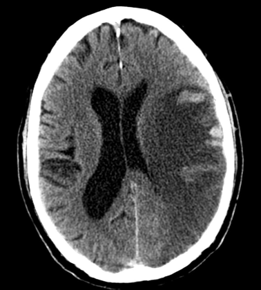
Interpret the following CT brain carefully.
Step-to-step interpretation :
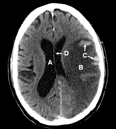
This diagram is at the level of the body of lateral ventricle ( A ). The left hemisphere shows a massive area of radiolucency in contrast to the normal density of the right cerebral hemisphere, suggestive of infarction in ischemic stroke ( Infarcted cells can absorb less X-ray i.e. more radiolucent ).
Noted that the infarcted area is roughly at the MCA territory i.e. the patient has left MCA occlusion. One of the essential learning point about ischemic stroke is that tPA, a common thrombolytic agent used in acute ischemic stroke, can no longer be given if there is hemorrhagic transformation from ischemic stroke ( because blood can leak if even the vessels get infarcted in later stage of ischemic stroke ).
In this case, we can see some whitish hemorrhages mixing with the infarcted area ( C ), indicating hemorrhagic transformation. There is a mild mid-line shift ( D ) due to cerebral edema after ischemic stroke. In summary, this is a left ischemic stroke in the MCA territory with a mild mid-line shift complicated by hemorrhagic transformation.


