Spontaneous Parenchymal Haemorrhage
Intra-ventricular extension of intra-cerebral haemorrhage
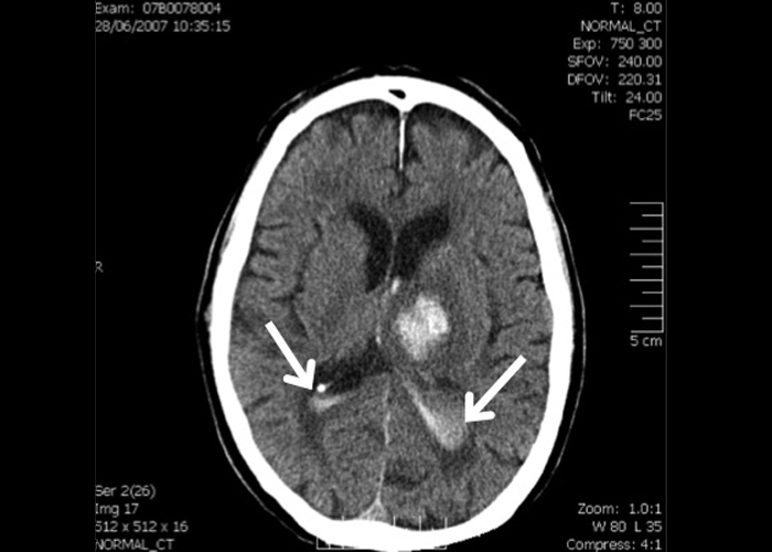
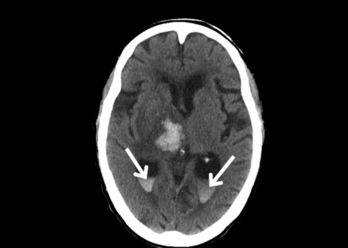
Occasionally, the intra-cerebral haemorrhage may extend into the ventricles ![]() (intra-ventricular extension) and these are most often seen in patients with haemorrhage involving the thalamus and caudate nucleus (Figures 1 and 2).
(intra-ventricular extension) and these are most often seen in patients with haemorrhage involving the thalamus and caudate nucleus (Figures 1 and 2).
Important acute management issues of patients presenting with spontaneous parenchymal haemorrhage includes: strict control of blood pressure as well as correction of any coagulopathies. Patient’s neurological status should be monitored closely as those with significant cerebral haemorrhage may result in coning and death. Approximately 10% of patients would also develop seizures as a result of the intra-cerebral haemorrhage. The patient should be investigated as to the underlying cause of the intra-cerebral haemorrhage and further vascular imaging (e.g. angiography) and a dedicated MRI scan to detect presence of underlying micro-bleeds should be considered in patients with a lobar haemorrhage. Whilst stable patients with small (<2cm), deep haemorrhages due to chronic hypertension could be managed medically, the Neurosurgical colleagues should be consulted for consideration of neurosurgical intervention if the haemorrhage is superficial, large and if the patient has worsening neurological status e.g. due to large clot volume causing significant mass effect, obstructive hydrocephalus etc.