| |

Spontaneous Parenchymal Haemorrhage

Intra-cerebral haemorrhage causing obstructive hydrocephalus

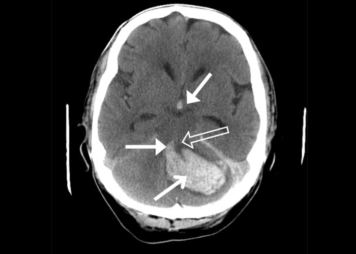
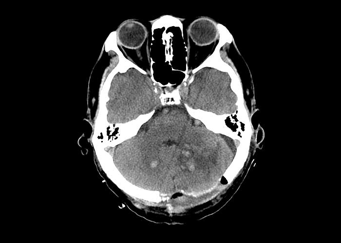
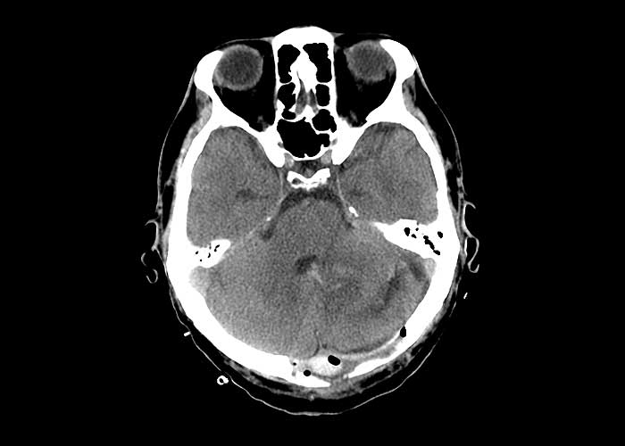
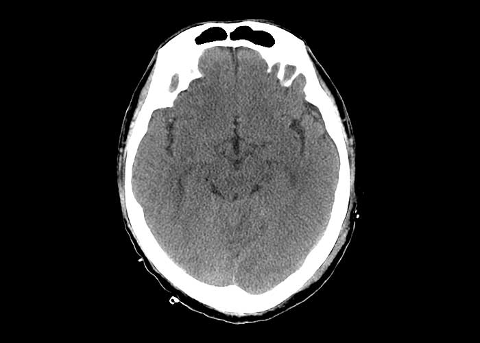
The first non-contrast CT shown here (Figure 1) is from a patient with large left-sided cerebellar haemorrhage arrow_1 with intra-ventricular extension arrow_4
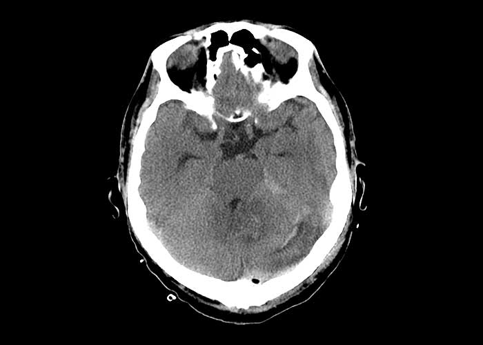
. The haemorrhage is causing significant mass effect on the 4th ventricle  arrow_5 
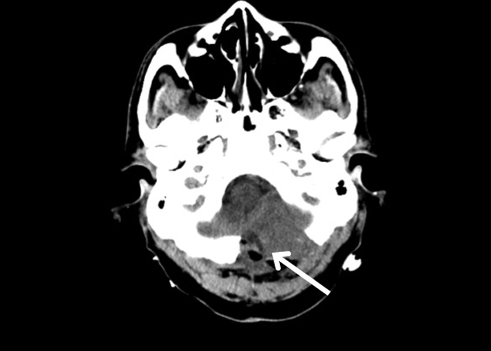
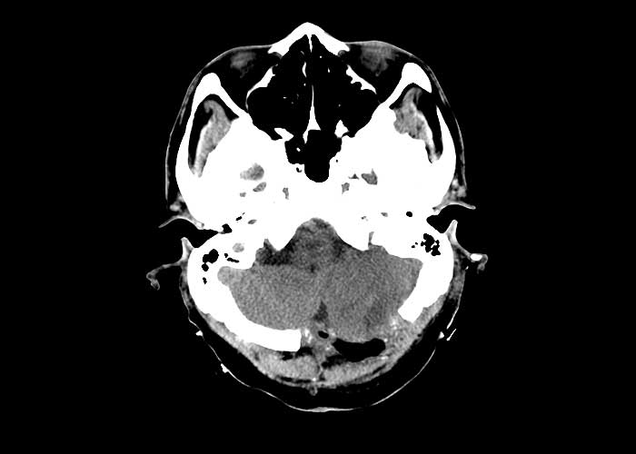
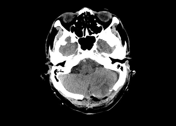
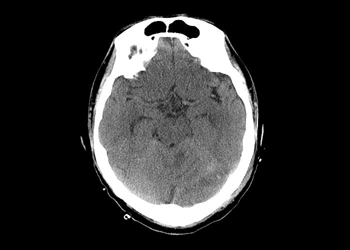
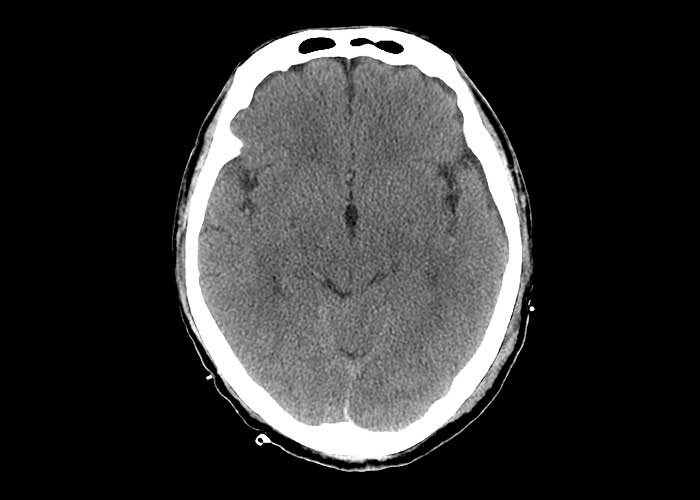
(resulting in obstructive hydrocephalus ) as well as the pons . The Neurosurgical team was consulted urgently and the patient was operated (Figures 7 to 12). A craniectomy was performed to relieve the intracranial pressure arrow_1 
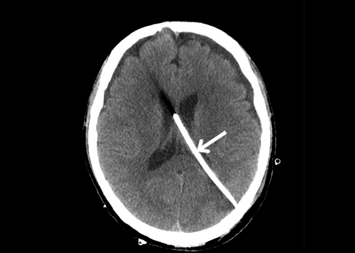
(a skull defect could be seen) (Figures 2 to 6), the cerebellar haematoma was extracted and an intra-ventricular drain was inserted arrow_1 (Figure 12).

Similar Posts

  • Frontotemporal Dementia

    Frontotemporal dementia is the most common cause of dementia in patients under the age of 65. There are three different subtypes: 1) behavioural-variant frontotemporal dementia,the most common subtype which is characterised by changes in personality and social conduct; 2) progressive nonfluent aphasia – a disorder of language output that may occur in the absence of…

  • Parkinson-plus Syndromes

    The Parkinson-plus syndromes include 1) multi-system atrophy, 2) progressive supra-nuclear palsy and 3) corticobasal degeneration. Multi-system atrophy Multi-system atrophy (MSA) is a rare adult-onset sporadic neurodegenerative disorder. There are two main subtypes, as identified by its dominating symptomatology. MSA-P is when parkinsonian symptoms predominate, and MSA-C if cerebellar symptoms dominate. These two terms have replaced…

  • Arterio-venous Malformations

    Cerebral vascular malformations can be classified by histopathology into 4 major types: Arterio-venous malformations (AVMs) Venous angiomas (developmental venous anomalies) Capillary telangiectasias and Cavernous malformations Arterio-venous malformations (AVMs) are congenital vascular lesions due to dysregulated angiogenesis. They are most often solitary but rarely could be multiple, in which case an underlying syndrome (e.g. hereditary haemorrhagic telangiectasia,…

  • Osmotic Demyelination Syndrome

    This is a set of cranial MRIs from a patient with osmotic demyelination syndrome, formerly called central pontine myelinolysis. Osmotic demyelination syndrome occurs when wide fluxes in serum sodium levels are induced by too rapid correction of hyponatremia. Oligodendrocytes, which forms the myelin sheaths, are particularly vulnerable to osmotic changes. If the osmotic stress is…

  • Dementia with Lewy Bodies

    Dementia with Lewy bodies is the second most common neurodegenerative dementia after Alzheimer’s disease, accounting for 15-20% of all cases. This form of dementia is due to an abnormal accumulation of Lewy bodies, which are spherical intra-neuronal protein aggregates consisting primarily of α-synuclein. Dementia with Lewy bodies is therefore considered as a synucleinopathy. Other synucleinopathies…