Ischaemic Changes of Different Anatomical Regions or Vascular Territories
Cerebellar infarct
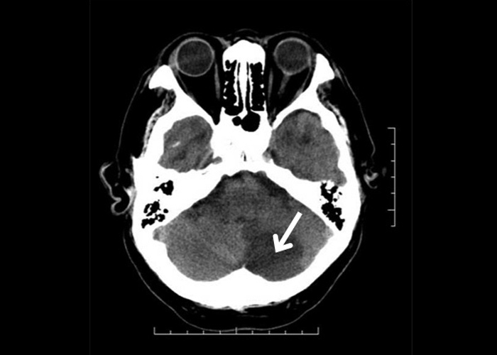
This non-contrast CT is from an elderly woman with atrial fibrillation who presented with an acute onset of left sided ataxia one month ago. The CT brain shows a hypodense lesion involving the left cerebellum ![]() (Figure 1). The area affected is supplied by the left posterior inferior cerebellar artery.
(Figure 1). The area affected is supplied by the left posterior inferior cerebellar artery.