
| |

Ischaemic Changes of Different Anatomical Regions or Vascular Territories

Infarction involving the subcortical territory: Basal ganglia infarct

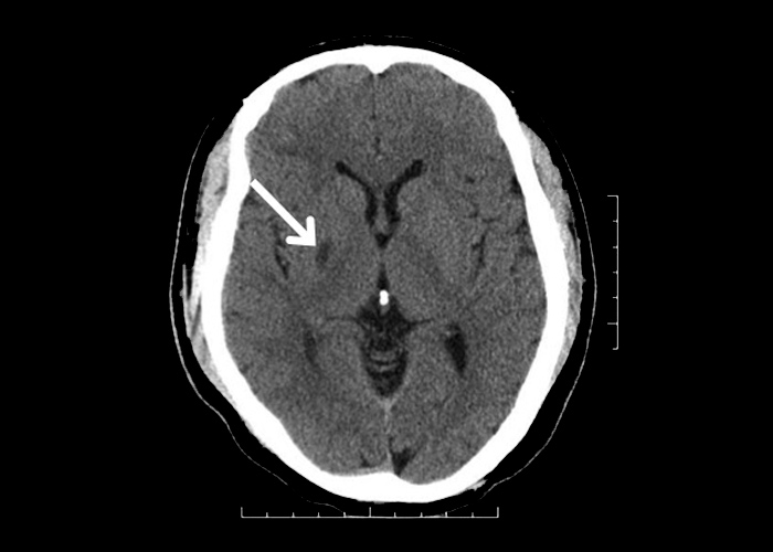
A small hypodense lesion is noted at the right lentiform nucleus in this non-contrast CT arrow_1 (Figure 1). This is a lacunar infarct and is usually due to small vessel occlusion as a result of chronic hypertension. Lacunar infarcts are defined as lesions that are ≤1.5cm in size on brain imaging.

Patients usually present with one of the 5 lacunar syndromes:

  1. Pure motor stroke
  2. Pure sensory stroke
  3. Mixed sensorimotor stroke
  4. Ataxic hemiparesis and
  5. Dysarthria-clumsy hand syndrome

This patient presented with acute onset of left sided weakness several years ago. There were no sensory signs nor any cortical or brainstem signs.
Similar Posts

  • | |

    Subarachnoid Haemorrhage

    Subarachnoid haemorrhages are where there is an extravasation of blood into the space between the arachnoid and pia. Subarachnoid haemorrhages often develop as a result of rupture of a cerebral aneurysm (most often arising from the circle of Willis or the middle cerebral artery bifurcation) or consequent to severe head trauma. Patients with subarachnoid haemorrhage secondary to aneurysmal…

  • Thymoma

    In this contrast thoracic CT scan of a middle-aged woman with newly diagnosed seropositive generalized myasthenia gravis, an abnormal thymic lesion (present in the anterior mediastinal compartment) could be seen  (Figure 1). The patient was referred to the Cardiothoracic Surgeons where she received a thymectomy via an open sternotomy. The thymic specimen was sent for histo-pathological…

  • Frontotemporal Dementia

    Frontotemporal dementia is the most common cause of dementia in patients under the age of 65. There are three different subtypes: 1) behavioural-variant frontotemporal dementia,the most common subtype which is characterised by changes in personality and social conduct; 2) progressive nonfluent aphasia – a disorder of language output that may occur in the absence of…

  • Autoimmune Encephalitis

    This cranial MRI is from a young woman who presented with low-grade fever, confusion, visual hallucinations and status epilepticus. Significant orofacial dyskinesias and autonomic dysfunction was also noted. Lumbar puncture was performed and cerebrospinal fluid (CSF) revealed mild lymphocytic predominant leukocytosis and slightly elevated protein. CSF glucose was within normal range and CSF for microbiological…

  • Obstructive Hydrocephalus

    These are the cranial CT scans from a patient with a recent left cerebellar infarct . Due to the significant cytotoxic oedema as a result of the cerebellar infarct, the fourth ventricle is being compressed , thus obstructing the normal CSF flow. The CT scan shows features of obstructive hydrocephalus and there is dilatation of the third ventricle  (now…