Ischaemic Changes of Different Anatomical Regions or Vascular Territories
Infarction involving the subcortical territory: Corona radiata infarct
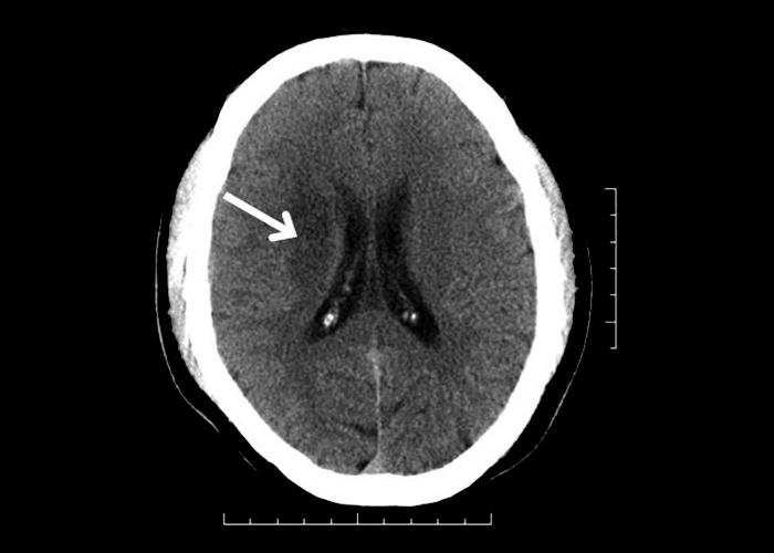
The first scan shown is a non-contrast CT demonstrating a hypodense lesion at the right corona radiata ![]() (Figure 1). The next scan is a MRI (diffusion weighted imaging (DWI) sequence) from another patient, also with an infarct involving the corona radiata. The scan shows evidence of “restricted diffusion” over this region
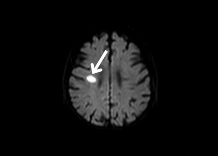
(Figure 1). The next scan is a MRI (diffusion weighted imaging (DWI) sequence) from another patient, also with an infarct involving the corona radiata. The scan shows evidence of “restricted diffusion” over this region ![]() (Figure 2). Both of these patients presented with an acute onset of left hemiparesis, slurring of speech and left facial weakness of an upper motor neuron lesion pattern.
(Figure 2). Both of these patients presented with an acute onset of left hemiparesis, slurring of speech and left facial weakness of an upper motor neuron lesion pattern.