Ischaemic Changes of Different Anatomical Regions or Vascular Territories
Cerebral infarction involving the anterior circulation
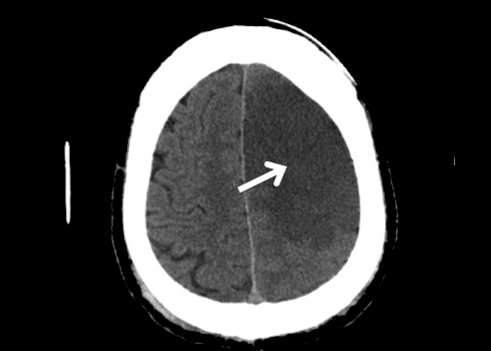
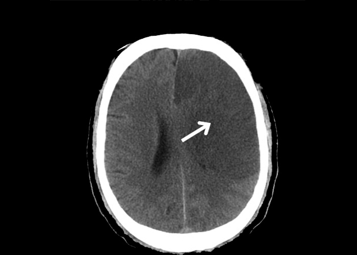
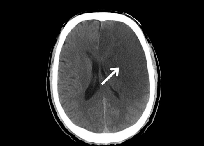
This non-contrast CT shows features suggestive of a subacute infarct at the left anterior cerebral artery territory and left middle cerebral artery territory ![]() (Figures 1-3). There is reduced grey-white differentiation and sulcal effacement at the cerebral cortex supplied by the left anterior cerebral artery and middle cerebral artery with a mild degree of mid-line shift to the right side due to cerebral oedema. Subsequent Doppler carotid ultrasound studies demonstrated that the patient had a complete occlusion of the left internal carotid artery resulting in the stroke.
(Figures 1-3). There is reduced grey-white differentiation and sulcal effacement at the cerebral cortex supplied by the left anterior cerebral artery and middle cerebral artery with a mild degree of mid-line shift to the right side due to cerebral oedema. Subsequent Doppler carotid ultrasound studies demonstrated that the patient had a complete occlusion of the left internal carotid artery resulting in the stroke.