Ischaemic Changes of Different Anatomical Regions or Vascular Territories
Cerebral infarction involving the middle cerebral artery territory: Acute middle cerebral artery territory infarct
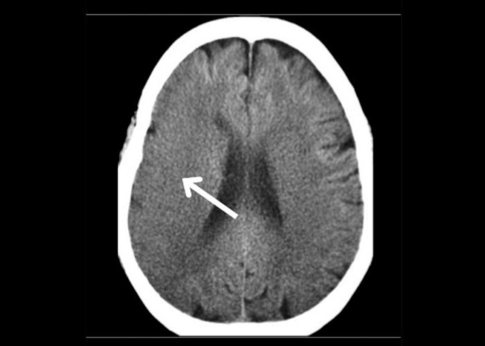
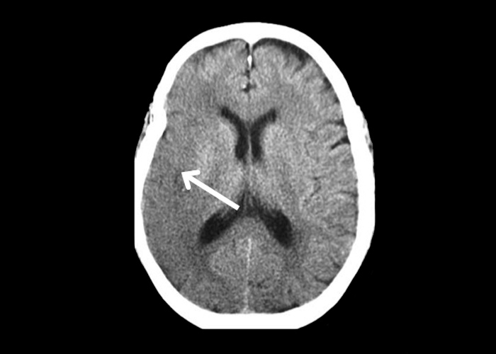
This set of non-contrast CTs is from a patient with an acute right middle cerebral artery infarct ![]() (Figures 1 and 2). There is reduced grey-white differentiation and sulcal effacement at the cerebral cortex supplied by the right middle cerebral artery. In addition, the right insular cortex as well as the lateral border of the right lentiform nucleus is less well demarcated compared to the left side.
(Figures 1 and 2). There is reduced grey-white differentiation and sulcal effacement at the cerebral cortex supplied by the right middle cerebral artery. In addition, the right insular cortex as well as the lateral border of the right lentiform nucleus is less well demarcated compared to the left side.